Lethal Dysrhythmias: VT, VF and Asystole
What it is
Ventricular tachycardia (VT), ventricular fibrillation (VF) and asystole stop effective ventricular pumping, so cardiac output and cerebral perfusion fall within seconds. Pulseless VT and VF are shockable: defibrillation depolarizes the whole myocardium so the sinus node can take over. Asystole and pulseless electrical activity are not shockable; they are treated with CPR, epinephrine and correction of the cause. The client's pulse, not the monitor alone, decides the treatment.
What the NCLEX focuses on
- When the monitor shows a lethal rhythm, assess the client first (responsiveness, breathing, central pulse) because a loose lead produces the same picture.
- Pulseless VT or VF: CPR, then an unsynchronized shock as soon as the defibrillator arrives, then 2 minutes of CPR before the next rhythm check.
- VT with a pulse and instability (hypotension, altered mental status, chest pain, signs of shock): synchronized cardioversion.
- Stable VT with a pulse: 12-lead ECG and an antiarrhythmic such as amiodarone.
- Asystole: high-quality CPR and epinephrine as soon as possible; never shock.
- Torsades de pointes is linked to a prolonged QT interval and hypomagnesemia and is treated with IV magnesium sulfate.
- Reversible causes (Hs and Ts): hypovolemia, hypoxia, acidosis, hypo- or hyperkalemia, hypothermia, tension pneumothorax, tamponade, toxins, thrombosis.
Priority nursing actions
- Check responsiveness, breathing and a carotid pulse for no more than 10 seconds.
- Call the emergency team and bring the defibrillator or AED.
- Start high-quality chest compressions.
- Defibrillate VF or pulseless VT as soon as possible, then resume compressions immediately.
- Give epinephrine and, for shock-refractory VF or VT, amiodarone as prescribed; search for reversible causes.
Numbers to know
| Item | Standard | Why it matters |
|---|---|---|
| Compression rate | 100 to 120/min | Maintains coronary perfusion pressure |
| Compression depth (adult) | At least 2 inches (5 cm), no more than 2.4 inches (6 cm) | Effective without injury |
| Compression-to-ventilation ratio | 30:2 without an advanced airway | Minimizes pauses |
| Pulse check / pause | No more than 10 seconds | Interruptions lower survival |
| Epinephrine | 1 mg IV or IO every 3 to 5 minutes | Raises coronary and cerebral perfusion |
| Amiodarone | 300 mg IV first dose, then 150 mg | For shock-refractory VF or pulseless VT |
| Compressor change | Every 2 minutes | Fatigue reduces compression quality |
Common exam traps
- Choosing to defibrillate asystole: wrong because there is no electrical activity to reset; CPR and epinephrine are the treatment.
- Choosing synchronized cardioversion for pulseless VT or VF: wrong because the device cannot find an R wave and the shock is delayed.
- Choosing IV amiodarone before cardioversion for VT with a weak pulse and BP 74/40 mm Hg: wrong because an unstable client needs synchronized cardioversion now.
- Choosing to stop compressions to look at the monitor: wrong because pauses lower coronary perfusion; rhythm checks occur every 2 minutes.
What to teach
- A client with an ICD carries the device card and tells every provider about it before procedures or MRI.
- After an ICD shock, call the provider; call 911 for more than one shock or for a shock with chest pain, dyspnea or fainting.
- Family members learn hands-only CPR and how to call emergency services.
Try 2 NCLEX-style questions on this topic
From the NCLEXPace question bank. Pick your answers, then check them inside NCLEXPace.
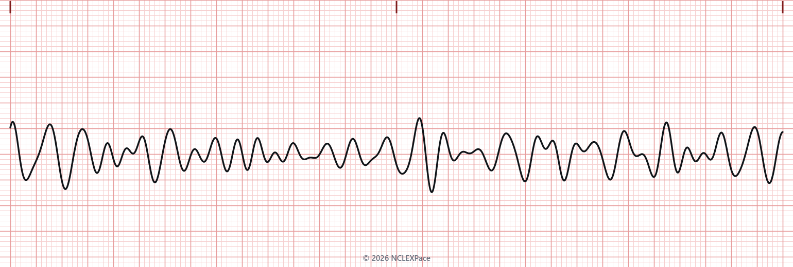
Select the best answer.
- A. Activate the emergency response team
- B. Prepare the defibrillator for an unsynchronized shock
- C. Begin chest compressions immediately
- D. Check responsiveness and a central pulse
Select the best answer.
- A. A trained unlicensed assistive personnel (UAP) performs chest compressions at 100 to 120 per minute.
- B. The respiratory therapist ventilates the client with a bag-mask device and oxygen.
- C. A nurse charges the defibrillator to deliver an unsynchronized shock.
- D. A nurse administers IV epinephrine according to the cardiac arrest protocol.
The correct answers, the reasoning for every option and 3,200+ more NCLEX questions are inside NCLEXPace. Create your account, then choose a plan to open them.
Create Account →Read more on this topic
Arrhythmias on the NCLEX: Every Rhythm With Its ECG StripMore tips on Medical-Surgical
Acute PancreatitisRecognize acute pancreatitis by pain and lipase, give fluids and pain control first, and catch hypocalcemia, shock and respiratory failure.Bowel Obstruction and NG Tube CareTell small from large bowel obstruction, spot strangulation early, and keep a decompression NG tube placed, patent and safe.Acute Kidney Injury vs Chronic Kidney DiseaseClassify AKI as prerenal, intrarenal or postrenal, follow its phases, and compare it with the staged, irreversible course of CKD.Sources
- Harding MM, Kwong J, Hagler D, Reinisch C. Lewis's Medical-Surgical Nursing: Assessment and Management of Clinical Problems. 12th ed. Elsevier; 2023; Chapter: Dysrhythmias.
- Wigginton JG, Agarwal S, Bartos JA, et al. Part 9: Adult Advanced Life Support: 2025 American Heart Association Guidelines for Cardiopulmonary Resuscitation and Emergency Cardiovascular Care. Circulation. 2025;152(suppl 2):S538-S577.
3,200+ NCLEX questions with think-like-a-nurse rationales, unfolding case studies and adaptive readiness exams. Start with free practice questions, then choose a plan when you are ready.
Create Account →