
Arrhythmias on the NCLEX: Every Rhythm With Its ECG Strip
Reading a rhythm strip is a five-step procedure, not pattern recognition: check the rate, whether it is regular, whether a P wave precedes every QRS, the PR interval, and the QRS width, where narrow is usually survivable and wide is usually urgent. The single distinction the exam tests hardest is which rhythms you shock and which you never do: defibrillate ventricular fibrillation and pulseless ventricular tachycardia, but never shock asystole or pulseless electrical activity.
Rhythm questions feel like pattern recognition and are actually a procedure. Learn the five-step read, then learn what each rhythm demands of you — and the one distinction that matters more than all the others: which rhythms you shock and which you never do.
The strips below are teaching diagrams drawn to show each rhythm's defining feature clearly. Real tracings have artefact and variation; use these to learn what to look for.
Step 1 — The five-step read
| Ask | What it tells you |
|---|---|
| 1. Rate? | Under 60, 60–100, or over 100 |
| 2. Regular? | Measure R to R. Irregularly irregular points straight at atrial fibrillation |
| 3. P wave before every QRS? | No P waves means it did not start in the sinus node |
| 4. PR interval? | Normal 0.12–0.20 s. Long, lengthening, or unrelated to the QRS points at a block |
| 5. QRS width? | Under 0.12 s is narrow — the beat came from above the ventricles. Wide means ventricular, and wide is usually the dangerous answer |
One shortcut carries a lot of weight: narrow is usually survivable, wide is usually urgent.
Sinus rhythms — the same rhythm at different rates
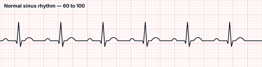
Everything present and in order: a P before every QRS, constant PR, narrow QRS, regular.
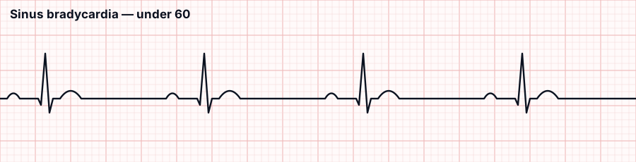
Sinus bradycardia. Normal in athletes and during sleep. Treat only if symptomatic — dizzy, hypotensive, chest pain, confused. Then atropine. Check the medication list first: beta blockers, digoxin and calcium channel blockers cause this.
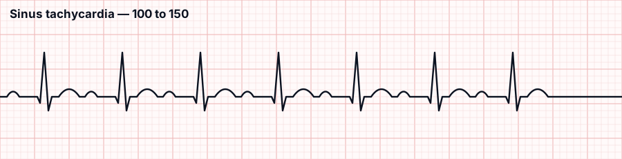
Sinus tachycardia. Almost never the problem — it is the response to one. Fever, pain, hypovolaemia, hypoxia, anxiety, blood loss. Treat the cause, not the rate. An option offering to slow this heart without finding out why is wrong.
Atrial rhythms

Atrial fibrillation. No P waves, a wobbling baseline, and R-R intervals that are irregularly irregular. The atria quiver rather than contract, so blood pools — and the real danger is not the rhythm, it is the clot.
- Stroke risk is the reason it matters. Anticoagulation is the priority intervention, not rate control.
- Rate control with beta blockers, diltiazem or digoxin.
- Before elective cardioversion of AF lasting over 48 hours, the client must be anticoagulated — shocking a fibrillating atrium back into contraction can launch a clot.
- Check an apical pulse for a full minute; a radial pulse under-counts because weak beats do not reach the wrist. That difference is the pulse deficit.
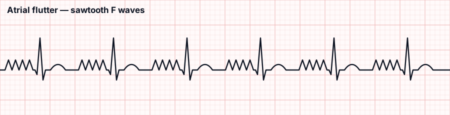
Atrial flutter. The sawtooth pattern is the whole diagnosis. Same clot risk and broadly the same management as atrial fibrillation.
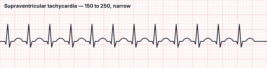
Supraventricular tachycardia. Very fast, regular, narrow, with P waves lost in the preceding T wave. Management escalates in a fixed order:
- Vagal manoeuvres — bear down, cough.
- Adenosine, given as a rapid IV push followed immediately by a rapid saline flush. Warn the client they will feel flushing and chest pressure, and expect a brief pause on the monitor — that is the drug working, not an arrest.
- Synchronized cardioversion if unstable.
Ventricular rhythms — where it gets dangerous
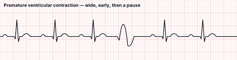
Premature ventricular contraction. One wide, bizarre, early beat, then a compensatory pause. Occasional PVCs are common and often benign. What makes them reportable:
- More than 6 per minute, or increasing in frequency
- Couplets (two in a row) or runs of three or more — that is ventricular tachycardia
- Multifocal — differently shaped, meaning several irritable sites
- Any PVC in the setting of an acute MI
First things to check: potassium and magnesium. Low levels of either provoke ventricular irritability, and correcting them is often the treatment.
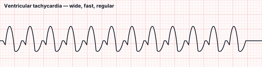
Ventricular tachycardia. Wide, fast, regular, no P waves. Everything depends on one question: does the client have a pulse?
| VT with a pulse | Pulseless VT |
|---|---|
| Antiarrhythmic (amiodarone); synchronized cardioversion if unstable | Defibrillate immediately and start CPR — treat it as ventricular fibrillation |
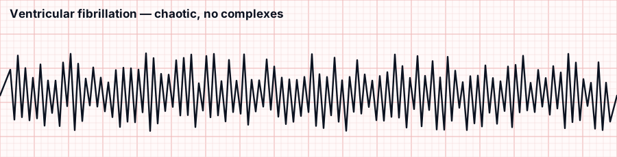
Ventricular fibrillation. Chaotic, no identifiable complexes, no cardiac output. The client is in arrest. Defibrillate immediately, high-quality CPR, epinephrine. Nothing else comes first.
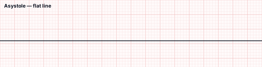
Asystole. Flat line. You do not shock this. CPR and epinephrine, and confirm it is real — check leads and connections, because a disconnected lead looks identical.
The distinction the exam tests hardest
| Shock it | Never shock it |
|---|---|
| Ventricular fibrillation Pulseless ventricular tachycardia | Asystole Pulseless electrical activity (PEA) |
| Defibrillate, then CPR | CPR and epinephrine; find and fix the cause |
And know the difference between the two kinds of shock:
| Defibrillation | Synchronized cardioversion | |
|---|---|---|
| Used for | VF, pulseless VT | Unstable but perfusing rhythms — AF, SVT, VT with a pulse |
| Timing | Immediate, unsynchronised | Timed to the R wave |
| Consent and sedation | No time — the client is unresponsive | Yes, the client is awake |
Safety for either: "clear" called aloud, nobody touching the bed or client, oxygen moved away from the field.
The AV blocks
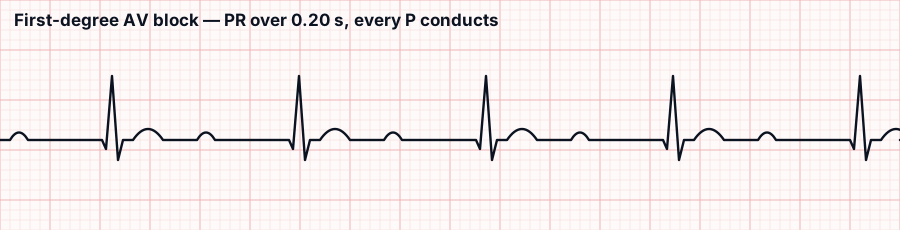
First degree. Every P conducts, but slowly — PR over 0.20 s and constant. Usually benign. Monitor, and review the rate-slowing drugs.
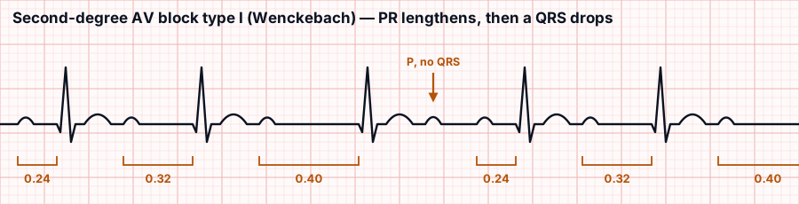
Second degree, type I (Wenckebach). The PR interval lengthens beat by beat until one QRS is dropped altogether — you can see the lone P wave with nothing after it. Usually benign; monitor and hold the offending drugs.
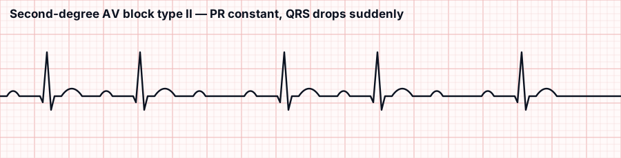
Second degree, type II. PR stays constant, then a QRS drops without warning. This one is serious — it progresses to complete block and it needs a pacemaker.
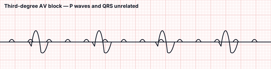
Third degree (complete). The atria and ventricles beat independently — regular P waves, regular QRS, no relationship between them. Pacemaker, and atropine often will not help because the problem is below the AV node.
| Block | The giveaway | Action |
|---|---|---|
| First degree | Long but constant PR | Monitor |
| Second degree type I | Longer, longer, longer, drop | Monitor |
| Second degree type II | Constant PR, sudden drop | Pacemaker |
| Third degree | P and QRS unrelated | Pacemaker |
NCLEX tip: Before anything else, ask whether the QRS is wide or narrow, and whether the client has a pulse. Wide plus no pulse means defibrillate. Flat line means never defibrillate. Those two facts answer more rhythm questions than memorising all fourteen strips.
Frequently asked questions
Common follow-up questions on Labs, Values & Diagnostics.
What are the five steps to read any rhythm strip?
Ask five questions: What is the rate? Is it regular, measuring R to R, since irregularly irregular points at atrial fibrillation? Is there a P wave before every QRS? What is the PR interval, normally 0.12 to 0.20 seconds? And how wide is the QRS, where under 0.12 seconds is narrow and came from above the ventricles, while wide means ventricular and is usually the dangerous answer.
Which rhythms do you shock and which do you never shock?
Shock ventricular fibrillation and pulseless ventricular tachycardia by defibrillating, then start CPR. Never shock asystole or pulseless electrical activity; those get CPR and epinephrine while you find and fix the cause. With asystole, confirm it is real by checking leads and connections, because a disconnected lead looks identical to a flat line.
What is the difference between defibrillation and synchronized cardioversion?
Defibrillation is used for VF and pulseless VT, delivered immediately and unsynchronised, with no time for consent because the client is unresponsive. Synchronized cardioversion is used for unstable but perfusing rhythms like AF, SVT, and VT with a pulse, timed to the R wave, and the awake client needs consent and sedation. For either, call "clear" aloud, keep everyone off the bed, and move oxygen away.
Why does atrial fibrillation matter and how is it managed?
In atrial fibrillation there are no P waves, a wobbling baseline, and irregularly irregular R-R intervals; the atria quiver rather than contract so blood pools, and the real danger is the clot, making stroke risk the reason it matters. Anticoagulation is the priority intervention, not rate control. Before elective cardioversion of AF lasting over 48 hours the client must be anticoagulated, and you check an apical pulse for a full minute.
Which AV blocks need a pacemaker?
First degree, with a long but constant PR, and second degree type I (Wenckebach), where the PR lengthens beat by beat until a QRS drops, are usually benign and monitored. Second degree type II, where the PR stays constant then a QRS drops without warning, is serious and needs a pacemaker. Third-degree complete block, where P waves and QRS are unrelated, also needs a pacemaker, and atropine often will not help.
3,000+ NCLEX questions with think-like-a-nurse rationales, unfolding case studies and adaptive mock exams: free to start, no card required.
Start Free Trial →